If you are citizen of an European Union member nation, you may not use this service unless you are at least 16 years old.
You already know Dokkio is an AI-powered assistant to organize & manage your digital files & messages. Very soon, Dokkio will support Outlook as well as One Drive. Check it out today!
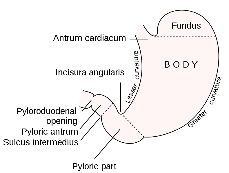
The stomach, which receives food from the esophagus, is located in the upper left quadrant of the abdomen (right below the diaphram). Its main function is to continue the breakdown of food, a process which begins in the mouth. The stomach is divided into the cardiac, fundic, body, and pyloric regions, and the lesser and greater curvatures are on the right and left sides, respectively, of the stomach.
I Anatomy of the stomach:
A) Basic Anatomy, starting at the superior end of the stomach:
a) Cardia: intersection point of the stomach and the esophagus (the cardiac orifice); the cardioesophageal sphincter (lower esophageal sphincter) separates the stomach from the esophagus
b) Fundus:the enlarged portion to the left and above the cardiac orifice; holds carbon dioxide created during the breakdown process and active in acid secretion
c) Rugae: folds lining the walls of the stomach. The rugae expand to allow more room for food without increased pressure in the stomach
d) Body, or Corpus: the body of the stomach is generally described as "C-shaped," formed by the greater and lesser curvature, but changes shape with the motions that churn and move the food towards the inferior end of the stomach and the pyloric sphincter
e) Pyloric sphincter: regulates the passage of chyme, or the semifluid mass of partly digested food, into the duodenum
B) Layers of the Stomach wall: the mucosal (innermost), the muscularis (middle) and the serosal (outermost)
Mucosal: The mucosal layer itself can be divided into three layers: the mucosa (the epithelial lining of the gastric cavity), the muscularis mucosae (low density smooth muscle cells) and the submucosal layer (consisting of connective tissue interlaced with plexi of the enteric nervous system).
Muscularis: The muscularis layer can also be divided into three layers: the longitudinal (the most superficial), the circular and the oblique. The longitudinal layer of the muscularis can be separated into two different categories: a longitudinal layer begins at the esophagus and ends in the body, and a longitudinal layer that originates in the corpus and spreads into the duodenum.
Serosal: The serosal layer is the serous membrane (the mesogastrium) that seals off the stomach from the surrounding organs.
II Physiology of the stomach:
a) Main Functions of the Stomach/Pyloric sphincter --
Storage. Because of its accordionlike folds (called rugae), the wall of the stomach can expand to store two to four liters of material. Temporary storage is important because you eat considerably faster than the digestive system can break down food and absorb nutrients.
Breakdown of food - Chemical and Physical. The stomach mixes the food with water and gastric juice (whose main components are the digestive enzyme pepsin (produced by the chief cells), hydrochloric acid (produced by parietal cells), intrinsic factor (essential for vitamin B12 absorption), and mucus) to produce a creamy medium called chyme. The gastric glands secrete the gastric juice; the hydrachloric acid and the enzyme pepsin break down large food particles by loosening the protein bonds in food. The HCl also kills most bacteria that may accompany the food, therefore protecting the body from invading bacteria. Once protein digestion begins, the stomach is protected by the layer of mucus secreted by the mucous cells.
In addition, the three layers of smooth muscles in the muscularis churn the contents of the stomach, physically breaking food down into smaller particles and moving the food/chyme towards the pyloric sphincter.
Controlled release. Movement of chyme into the small intestine is regulated by a valve at the end of the stomach, the pyloric sphincter.
III Common Illnesses of the Stomach and Pyloric Sphincter:
1. Gastritis: an inflammation of the stomach lining. The main acute causes are poor diet, excessive alcohol consumption, and/or prolonged use of drugs such as asprin or ibuprofen. Some cases of gastritis develop after major surgery, traumatic injury, burns, severe infections, or those who have had weight loss surgery (resulting in the banding or reconstruction of the digestive tract).
Treatments:Acute gastritis caused by NSAIDs or alcohol may be relieved by stopping use of those substances. Chronic gastritis caused by H. pylori infection is treated by eradicating the bacteria. Most gastritis treatment plans also incorporate medications (antacids such as Maalox and Mylanta; acid blockers such as Tagamet, Zantac, Axid or Pepcid), that help reduce the amount of acid your stomach produces or try to neutralize stomach acid in order to reduce signs and symptoms.
2. Pyrosis: commonly known as heartburn because the area of pain is felt near the heart; occurs when stomach acid backs up into the esophagus (reflux). Can be caused by smoking, pregnancy, certain foods, alcohol and some medications.
Treatments: If the pyrosis is caused by smoking or medications, stop use of these immediatly. Smoking has harmful effects on the salivary function which helps to clear the esophagus from the acids, while some medications relax the Lower Esophageal Sphincter (LES), allowing stomach contents to reflux back up into the esophagus. Pyrosis can also be treated with antacids/acid blockers, much like gastritis. Another treatment, proton pump inhibitors (PPIs), consists of a group of prescription medications that prevent the release of acid in the stomach and intestines. Doctors prescribe PPIs to treat people with frequent pyrosis, ulcers of the stomach or intestine, or excess stomach acid.
Pyrosis is common during pregnancy; the placenta produces the hormone progesterone, which relaxes the cardioesophageal sphincter, allowing gastric acids to seep back up. Progesterone also slows down the wavelike contractions of your esophagus and intestines, making digestion sluggish. Later in pregnancy, the growing baby crowds your abdominal cavity, pushing the stomach acids back up into the esophagus.
3. GERD, or Gastroesophageal reflux disease: this is the frequent occurrence of reflux due to an incompetent lower esophageal sphincter, which allows acid to back up into the esophagus and can damage the esophagus over time. Treatment includes acid blockers and/or antacids, and sometimes surgery to repair damage done to the esophagus and valve by the reoccurring reflux.
4. Gastric (Peptic) Ulcers: occurs when the stomach lining is weakened, by the H. pylori bacteria, certain diseases, or nonsteroidal anti-inflammatory drugs (NSAIDs) such as aspirin and ibuprofen. The weakened wall is rendered defenseless against the stomach's own acid; the acid breaks down part of the stomach wall, leaving an open sore equal to or greater than 0.5 cm in width. Gastric ulcers may or may not become malignant. Treatment is usually similar to the treatments for pyrosis if the ulcer is caused by damage to the stomach wall; medications are prescribed to decrease the amount of acid in the stomach and allow the ulcer to heal. If the ulcer is caused by theH. pylori bacteria, then antibiotics will be prescribed to kill the bacteria.
5. Stomach Cancer: the formation of cancer cells in the stomach lining, usually occurring after an H. pylori bacterial infection (gastric ulcers) or presence of benign polyps, gastritis, prolonged exposure to salt and cigarette carcinogens; cause can also be related to genetics.
Metastasis occurs in 80-90% of individuals with stomach cancer, with a six month survival rate of 65% in those diagnosed in early stages and less than 15% of those diagnosed in late stages.
Diagnosis:
Fecal Occult Blood Test: This laboratory test is used to determine the presence or absence of hidden (occult) blood in the stool. The occult blood test is performed because stomach cancer sometimes causes bleeding that can't be seen. The presence of blood in the stool by itself is not diagnostic of stomach cancer. Other conditions can cause occult blood, such as eating meat within a day or so of the test.
Upper Gastriointestinal Series: The upper GI series involves taking x-rays of the esophagus and stomach after drinking a harmless solution of barium, a dye that makes the stomach easier to see on x-rays. This test, also called the barium swallow, outlines the stomach, helping the doctor or radiologist to locate any abnormal areas. During the test, the doctor may pump air into the stomach to make suspicious areas easier to see.
Endoscopy:An endoscope is a thin tube with a tiny camaralike end. During endoscopy (sometimes called gastroscopy), this tube is passed through the mouth and esophagus into the stomach for a direct visual examination. When the endoscope is in place, the doctor can see directly into the stomach. If an abnormal area is seen, a biopsy (seen below) can be performed to determine the presence or absence of cancer cells in the suspect tissue.
Treatments:
Surgery: total or partial gastrectomy, where either a part of the stomach or the whole stomach is removed. After a partial gastrectomy, the remaining portion of the stomach is connected to the esophagus or the small intestine, depending on which part of the stomach was removed; after a total gastrectomy, the esophagus is connected directly to the small intestine.
Chemotherapy: Chemotherapy is called a systemic treatment because when the drug enters the bloodstream, it travels throughout the body and can kill cancer cells outside the stomach. Chemotherapy drugs act either by destroying tumor cells or by preventing them from multiplying. When chemotherapy is used to treat stomach cancer, the drugs are usually prescribed in groups of three or four, called a protocol.
Radiation Therapy: Radiation therapy uses either an external machine to radiate cancer cells from outside the body (external radiation therapy), or brachytherapy (internal radiation therapy), which uses materials called radioisotopes that are introduced into the body through thin plastic tubes. This method enables the radiation to be guided directly to the area where the cancer is found.
Biological Therapy, or Immunotherapy: an experimental treatment where the body's own natural defenses are used to destroy cancer cells
This is your Sidebar, which you can edit like any other wiki page.
This Sidebar appears everywhere on your wiki. Add to it whatever you like -- a navigation section, a link to your favorite web sites, or anything else.


Comments (2)
csnanatomy said
at 2:56 pm on Apr 2, 2009
Can you show video of stomach digestion?
Hayley McPhedran said
at 10:58 am on Apr 3, 2009
I've been looking at upper digestive system colonoscopies, but I think I could find a stomach digestion video.
You don't have permission to comment on this page.