Pancreas,liver, gallbladderAnatomy of the Pancreas
The exocrine pancreas (regulates digestion) consists of acinar and duct cells.
Acinar cells: produce digestive enzymes and constitute the bulk of the pancreatic tissue; organized into grape-like clusters that are at the smallest termini of the branching duct system.
Ducts: add mucous and bicarbonate to the enzyme mixture; form a network of increasing size, culminating in main and accessory pancreatic ducts that empty into the duodenum.

Pancreatic Secretions
The pancreas aids in chemical digestion by producing an alkaline solution rich in bicarbonate as well as several enzymes. The bicarbonate neutralizes the acidity of chyme and acts as a buffer (pH). Among the pancreatic enzymes are trypsin and chymotrypsin, proteases secreted into the duodenum in inactive forms. In a chain reaction similar to activation of pepsin, they are activated when safely located in the extracellular space within the duodenum.
Hormone-secreting cells make up only 1-2% of the mass of the pancreas. Other cells in the pancreas produce and secrete bicarbonate ions and digestive enzymes. These secretions are released into small ducts that empty into the pancreatic duct, which leads to the small intestine. Thus, the pancreas is both an endocrine gland and an exocrine gland with functions in the endocrine and digestive systems.


Diseases of the Pancreas
Pancreatitis: inflammation of the pancreas caused when the digestive enzymes from the exocrine pancreas become activated inside of the pancreas, instead of in the duodenum, and start “digesting” the pancreas itself. It usually presents with abdominal pain and can cause nausea and vomiting.
The most common cause of acute pancreatitis is blockage of the pancreatic duct by gallstones. Acute pancreatitis often presents with raised levels of pancreatic enzymes in the blood, which can circulate to other body organs, causing shock and organ failure. Other causes can include biliary tract disease (obstruction, gallstones or sludge), alcohol excess, physical trauma to the abdomen, hyperlipidemia and hypercalcemia.
Chronic pancreatitis is characterized by chronic or persistent abdominal pain that develops gradually, often resulting in slow destruction of the pancreas and can lead to other problems such as pancreatic insufficienty, bacterial infections, and type 2 diabetes. The main causes are gall bladder disease (ductal obstruction) and alcoholism. Other causes include cystic fibrosis, hypercalcemia, hyperlipidemia, some drugs, and autoimmune conditions.
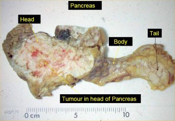
Pancreatic Cancer: (upper photos) fourth leading cause of cancer death in the United States (32,000 deaths annually). Risks include smoking, age, gender (more common in men), dietary factors, and chronic pancreatitis. Most (95%) pancreatic cancers are adenocarcinomas, developing in the exocrine tissues (predominantly ductal epithelium). Roughly 75% of pancreatic carcinomas occur at the head of the pancreas. Pancreatic cancer is difficult to detect in the early stages because symptoms are either absent or nonspecific: abdominal pain, nausea, loss of appetite, and sometimes jaundice.
Pancreatic Insufficiency: inability of the pancreas to produce and/or transport enough digestive enzymes to break down food in the intestine and allow its absorption. It typically occurs as a result of progressive pancreatic damage - damage that may be caused by a variety of conditions. It is most frequently associated with cystic fibrosis in children and with chronic pancreatitis in adults; it is less frequently but sometimes associated with pancreatic cancer.
The Endocrine Pancreas
Type 1 Diabetes/Insulin-Dependent Diabetes: autoimmune disorder in which the immune system destroys the beta cells (control the release of insulin into the blood) of the pancreas
Type 2 Diabetes/Non-Insulin-Dependent Diabetes: target cells fail to respond normally to insulin. Insulin is produced, but target cells fail to take up glucose from the blood, and blood glucose levels remain elevated.
Bile Production by the Liver
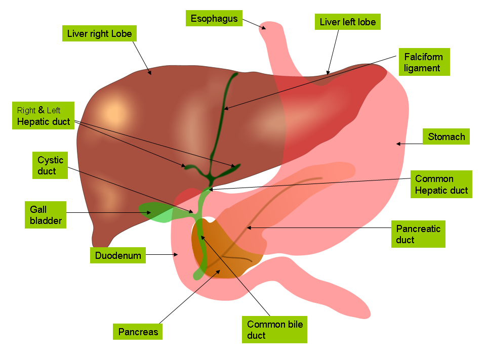
Digestion of fats and other lipids begins in the small intestine and relies on the production of bile, a greenish yellow, thick, sticky fluid mixture of substances that is made in the liver. Bile contains bile salts, which act as detergents (emulsifiers) that aid in digestion and absorption of lipids (cholesterol, fats, fat-soluble vitamins) in the intestine. Bile is stored and concentrated in the gallbladder. As digestion of food occurs in the stomach and small intestine, the gallbladder releases bile through a tube called the common bile duct. This duct connects the gallbladder and liver to the small intestine.
The liver has many vital functions in addition to bile production. It also breaks down toxins that enter the body and helps balance nutrient utilization. Bile production itself is integral to another task of the liver: the destruction of red blood cells that are no longer fully functional. In producing bile, the liver incorporates some pigments that are by-products of red blood cell disassembly (bilirubin). These bile pigments and cholesterol are then eliminated from the body with the feces.
The Anatomy and Major Functions of the Liver
Anatomy
The liver is the body's largest internal organ and one of its most important organs as well. It weighs around 1.4 kg, is a pinkish-brown color, and can be found just below the diaphragm in the abdominal cavity. The liver is divided into four different lobes: the left anatomical lobe, the right anatomical lobe, the caudate lobe, and the quadrate lobe. All four of these lobes are divided up by ligaments present in the visceral peritoneum that envelops the liver except at the bare area, where the liver attaches to the diaphragm. (**Note: the"ligaments" mentioned have no relation to the ligaments present in joints. They simply serve as landmarks). In between the quadrate and caudate lobes is the porta hepatis, a depression where the hepatic portal vein, the hepatic artery, and the common bile duct all attach to the liver. The branches of the artery, vein, and duct divide the liver into its right and left parts.
http://www.youtube.com/watch?v=JEgWR2oo4_M
http://www.youtube.com/watch?v=T3Rw7T9cVuQ
^^Helpful videos about the anatomy of the liver

Functions
Our bodies depend greatly on the liver to perform a variety of functions that are vital to our survival. First the liver detoxifies the blood, removing harmful substances from it. This detoxification helps fight infections and removes harmful bacteria. The liver also processes all the blood that leaves the intestines and stomach and breaks down the drugs and nutrients that are present in the bloodstream, turning them into substances that can be more easily used by the body. Another major function of the liver is the production of bile, which carries away the by-products that resulted from the breakdown of substances during blood detoxification from the intestines and stomach. The liver then sends the by-products to be excreted from the body. Other notable functions of the liver are the regulation and productions of certain proteins, the storage of sugar, and the digestion of fats from food.
http://www.nottingham.ac.uk/nursing/sonet/rlos/bioproc/liveranatomy/index.html
^^This website has a very informative video. Click on the link, look at the right side of the page, and click "start". There are several videos so make sure to watch all of them.
Diseases of the Liver
Hepatitis
Hepatitis is the swelling of the liver that causes it not to function correctly. Usually hepatitis is caused by viruses. These viruses are the factors that determine whether a person has hepatitis A, B, or C. Although viruses are the main causes of hepatitis, drug and alcohol abuse can also play a role in increasing a person's chance of contracting the disease. In rare cases, the hepatitis occurs when the body attacks its own tissues. A person suffering from any form of hepatitis can have any of these symptoms: loss of appetite, nausea, vomiting, diarrhea, dark-colored urine, pale colored bowel movements, stomach pain, or jaundice.
Hepatitis A (HAV)- HAV is a type of hepatitis that occurs when the A virus infects the liver causing swelling but rarely resulting in long term damage. In most cases, the liver heals on its own within a few weeks and sometimes symptoms are not present. HAV is commonly spread in four different ways: 1.) contaminated drinking water, 2.) not washing you hands after changing a diaper, 3.)sexual contact (includes oral or anal), 4.) when an infected person does not wash their hands after going to the bathroom and then comes in contact with food that is later consumed by another person.
.jpg)
Hepatitis B (HBV)- HBV is spread from an infected mother to her child during pregnancy, through bodily fluids such as blood and semen and can be short term or chronic. If a person suffers from chronic HBV, their liver can scar, resulting in even further complications. A blood test is necessary for a doctor to diagnose HBV and there is a vaccine available.

Hepatitis C (HCV)- HCV is transmitted in the same ways as the HBV virus and is the most serious of the three types of Hepatitis. The symptoms of HCV may not appear for years and a blood test i s necessary to diagnose it. There is not cure for HCV and it is a chronic condition and sometimes, a liver transplant is necessary.
Cirrhosis
Cirrhosis is a condition where the liver begins to malfunction due to chronic injury. As healthy tissue is replaced with scarred tissue, the liver's ability to allow blood flow, remove toxins/bacteria, make proteins that regulate blood clotting, process nutrients, control infections, and produce bile become impaired. There are a variety of ways a person can develop cirrhosis but the primary causes are extreme alcohol abuse and Hepatitis C. Symptoms of cirrhosis are fatigue, weight loss, nausea, vomiting, loss of appetite, itching, abdominal pain, spider-like blood vessels under the skin. If a patient is suffering from any of these symptoms, and MRI, ultrasound, or CT scan is typically performed to diagnose if the patient has cirrhosis.


Hepatocellular Carcinoma (HCC)
Hepatocellular Carcinoma is a specific type of liver cancer and amounts for 80%-90% of all liver cancers. Unlike most types of liver cancers, hepatocellular carcinoma originates within the liver. Many other types of liver cancers spread to the liver after first attacking the colon, stomach, or breasts. Often times, people suffering from cirrhosis end up developing hepatocellular carcinoma because alcohol abuse plays a major role in the death of hepatocytes (liver cells). If a person has chronic hepatitis B or hepatitis C, their chances of being diagnosed with hepatocellular carcinoma greatly increase. Cases of HCC are fairly low in the United States but are extremely high in Asia and Africa. If a patient is suspected of having HCC, an ultrasound or CT scan should be done immediately.
The Liver's Role in Cholesterol Production
The liver is responsible for producing about 80% of the cholesterol within the human body. As you digest, the liver takes in the cholesterol from the the blood stream that is produced by the food consumed and absorbs the triglycerides it needs. After the cholesterol is absorbed, it is packed with fats and proteins which later form lipoproteins. The lipoproteins are then distributed throughout the body. If an insufficient amount of dietary cholesterol is being consumed, the liver produces its own cholesterol. If the liver was not able to produce cholesterol, then vegetarians and vegans would not be able to obtain enough cholesterol and they would die. Cholesterol is needed in order to strengthen cells and aid in the production of hormones such as estrogen. However, over consumption of dietary cholesterol can have negative affects on the human body so it is important to find a healthy balance of cholesterol intake.
Anatomy and Physiology of the Gallbladder
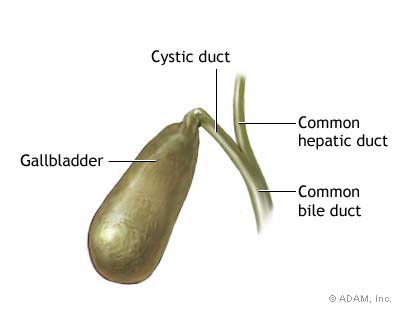
The gallbladder is a small, pear-shaped sac located beneath the liver. When bile is needed, as when people eat, the gallbladder contracts, pushing bile through small, tubelike passages (cystic duct and common bile duct) into the small intestine. The biliary tract consists of small tubes (ducts) that carry bile from the liver to the gallbladder and then to the small intestine for this purpose.

Diseases of the Gallbladder
Cholecystitis: (above right photo) inflammation of the gallbladder (sometimes thickening of the wall) that usually develops when an individual has gallstones, rocklike chemical deposits that form inside the gallbladder. Bile becomes trapped in the gallbladder if the cystic duct is blocked by a gallstone, which can lead to bacterial infection and subsequent inflammation of the gallbladder itself from the chemicals in the trapped bile.
Acute cholecystitis: sudden inflammation of the gallbladder causing abdominal pain
Chronic cholecystitis: damage to the walls of the gallbladder lead to a thickened, scarred gallbladder which ultimately can shrink and lose its ability to store and release bile
Biliary colic: steady or intermittent ache in the upper abdomen under the right side of the rib cage occurring when something blocks the normal flow of bile from the gallbladder. Gallstones are the most common reason for biliary colic. When a gallstone blocks either of the ducts, the normal flow of bile into the intestine is disrupted and the muscle cells in the bile duct contract vigorously to try to move the stone, causing the pain of biliary colic. The flow of bile can also be blocked by any of the following:
-gallstones that pass out of the gallbladder into ducts
-disorders of the pancreas, which can narrow the bile ducts that pass through the pancreas
-tumors in the pancreas or bile ducts
-infestation by parasites (in Asia)
Gallbladder Polyps: growths that protrude from the lining of the inside of the gallbladder that can be, but are rarely cancerous. Most gallbladder polyps are benign (indicated generally by a size of less than 1 cm) and require no treatment, however, those that are larger than 1 cm may be cancerous and require surgical removal of the gallbladder (cholecystectomy).
Comments (1)
csnanatomy said
at 2:55 pm on Apr 2, 2009
Looking good!
You don't have permission to comment on this page.